One hundred secrets and one "sparse" are difficult to recover! Don't let osteoporosis turn into compression fractures

Is it possible to have a “compression fracture” even without falling? Physiotherapist: Sudden low back pain and back pain are warning signs!
“Therapist, my mother started complaining about back pain in the middle of the night yesterday and has been unable to sleep well. Can you help her take a look?”
“No problem. Let me confirm first: Did Mother Qiu get up to go to the toilet last night? Did she fall?”
“Maybe. She usually gets up once or twice a night, but she said she didn’t fall down yesterday.”
“Based on the pattern of pain and your mother’s age, it looks like a compression fracture. I recommend taking an X-ray for follow-up confirmation.”
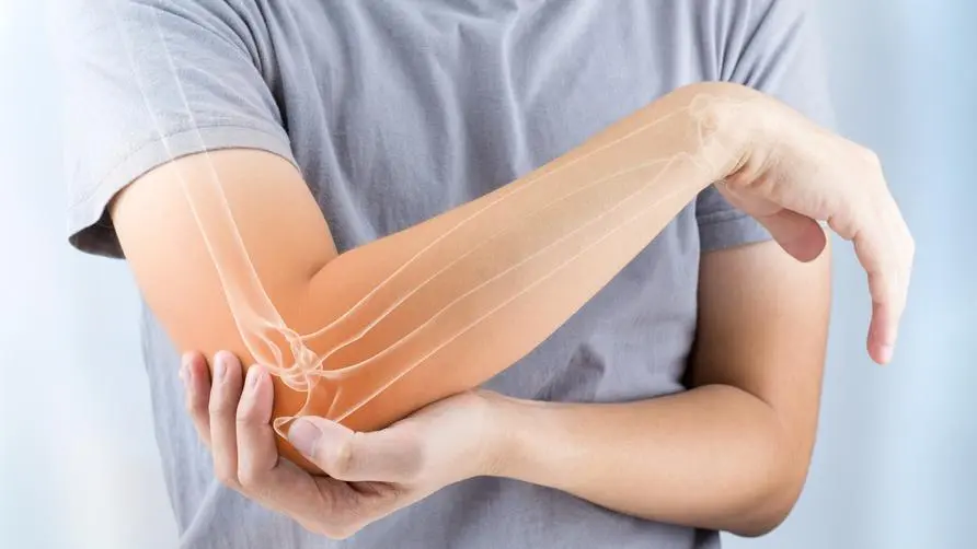
Osteoporosis is a silent disease. Many people may think that as long as they do not fall, then “bone loss” will not have a big impact on people. In fact, if there is insufficient bone in the body, it may cause back pain and shortening of height. Or changes in the shape of the spine such as stooping or hunching, or even more people may not realize that the bones have become fragile due to bone loss, so they do not know that a compression fracture has occurred until they experience pain in the hip joint, spine, wrist, etc.
What are the symptoms of spinal compression fractures? Seek medical attention immediately if your height “suddenly becomes shorter”
If a compression fracture occurs in the spine, it may cause severe back pain and difficulty in turning over, especially when sitting or standing upright. In clinical experience, it is common for some patients with severe osteoporosis to suffer compression fractures of the thoracic or lumbar vertebrae because they do not control the strength to sit down and sit heavily on a chair or bed. Of course, accidents such as falls, falls from heights or car accidents are also common causes.
If we look into the shape of the spine, each vertebral body is flat and cylindrical. If a force perpendicular to the vertebral body causes the vertebral body to collapse, a compression fracture will occur. On X-ray, it looks like a squashed scone. Even if they do not suffer from compression fractures, people with osteoporosis may still experience symptoms such as height shrinkage and limited spinal mobility.
Treatment of spinal compression fractures? If “1 training” is not done well, it may be more difficult to get better.
Currently, the commonly used treatment methods are brace fixation and bone cement surgery:
Bone cement surgery (bone cement grouting pyramidoplasty): This surgery is a minimally invasive surgery that uses a fine needle with a diameter of about 2-3 mm to inject bone cement (PMMA) into the fractured vertebra under image guidance. in the body to increase the strength and stability of the vertebral body and prevent it from further collapse.
Back brace fixation: Regardless of whether they undergo surgery or not, patients must wear a brace for 3-6 months to reduce the movement of the spine. During the period of using the brace, it is highly recommended to undergo rehabilitation therapy at the same time to avoid extending degenerative problems. .
Rehabilitation at this time will focus on core muscle training to maintain muscle status and prevent the patient from becoming weak in the trunk muscles due to long-term use of a back brace or lying completely flat in bed, making the patient more susceptible to subsequent injuries. In addition, in order to take into account the injury of spinal compression fracture, the core muscle group training at this time will avoid upright spine, so it is mostly performed in a lying down or four-legged kneeling position. For example: When lying on your back, do leg movements at the same time and breathe in conjunction.
How to prevent compression fractures? Neither bone preservation nor muscle gain can be neglected.
The basic prevention of compression fractures can start from two aspects: “preserving bone” and “increasing muscle strength.” Even in the face of osteoporosis caused by increasing age and hormonal changes after menopause, it can be effectively dealt with. The first is to preserve bone: a complete nutritional intake, such as calcium, magnesium, vitamin D or K, can help build bone defense and reduce the risk of osteoporosis.
In addition, common high-calcium foods include tofu, dairy products, kelp, black sesame seeds, etc. As for supplementing vitamin D, in addition to eating mushrooms, black fungus, egg yolks, and salmon, because the vitamin D content in food is very limited, it is generally recommended to let the body synthesize it by itself in the sun. On this basis, it is even more necessary to avoid unhealthy habits such as smoking, drinking alcohol, and staying up late so that these nutrients can be successfully retained in the body.
The third is “building muscle strength”. Weight-bearing exercises can not only keep bones dense, but also maintain muscle strength and reduce the risk of falls and fractures. In addition, increasing muscle mass can also increase metabolism, improve cardiopulmonary function, and fight aging. Moderate exercise also improves balance and coordination, reduces the risk of falls, improves muscle strength and physical function, and promotes overall health. However, attention should be paid to the intensity and method of exercise in a gradual manner to avoid the occurrence of sports injuries.
Make good use of bone density testing and osteoporosis medications to avoid injuries and long-term rehabilitation
Bone density examination is the most direct way to assess bone health! Early detection of bone quality in the lumbar spine and hip joints that are susceptible to osteoporosis can help prevent fractures and provide more accurate results than ultrasound examinations. Use low-dose X-rays such as DEXA (dual-energy X-ray absorptiometry) to scan bones, measure the extent to which X-rays penetrate the bones, and calculate bone density. A bone density measurement with a T-score less than or equal to -2.5 is diagnosed as osteoporosis.
However, because the DXA machine is larger than a single bed and is limited in the examination space, a quantitative ultrasound bone density detector (quantitative ultrasound, QUS) is sometimes used for measurement. If the measurement site is the heel, a quantitative ultrasound bone density detector is used. It is less accurate and is usually used as a preliminary screening tool. When medical staff confirm that a patient has osteoporosis, he or she will be given different medications based on gender and age to maintain bone mass.
Taking women as an example, in the early stage, if they are younger than 60 years old and have just stopped menstruating for less than ten years, they will be adjusted with hormonal therapy. Subsequently, according to changes in age, medications to increase bone formation and inhibit bone loss may be considered. Commonly used drugs include “Ariyi”, “Gylicon”, “Baokali”, etc. For people who have inconvenient return visits and are prone to forgetting to take medication, doctors usually recommend injections rather than oral medications.
It is not easy to rebuild bone density, and it is difficult to restore it once it is lost, but we can still slow down the rate of bone loss and avoid the risks caused by bone loss; and the best way to prevent osteoporosis is to pass it through early Bone density checks to ensure the current bone condition and maintain good exercise habits will save us from injuries and the hard work of rehabilitation. I came to Zhonglun International Clinic this month. After the doctor’s evaluation during the registration, I can do an on-site bone density test (Lunar iDXA), which will give my mother the encouragement of love and the strength of my family’s love!
Extended reading: Is osteoporosis not just for the elderly? She took a test and found that her bone density was “lower than her mother-in-law’s”! Experts reveal 7 extremely high-risk groups with bone loss like a house of sand in the sea! The 50-year-old woman had an artificial hip joint but it came loose. It turned out to be rheumatoid arthritis that caused the bone loss.