Are you afraid of radiation during ultrasound for heart disease? Can't a family member take a test if she's pregnant? Cardiologist: "New technology" does not need to worry about radiation problems

Ms. Huang, 70, went to the cardiology clinic because she had difficulty breathing, wheezing and chest tightness when climbing stairs. Ms. Huang had obvious symptoms of angina pectoris, which was suspected to be caused by coronary artery disease, so she was advised to undergo further examination. Her general cardiac ultrasound was approximately normal except for mild diastolic dysfunction and valvular regurgitation. The doctor suggested that she undergo a nuclear medicine examination to evaluate whether there is ischemia in the myocardium. However, because her daughter-in-law was just pregnant, she was afraid that it would affect the child, so she provided Ms. Huang with two options, including exercise electrocardiography and stress ultrasound plus deformation. analyze.
After discussion, Ms. Huang agreed to undergo a stress ultrasound examination. The results showed that myocardial ischemia was indeed present, and a cardiac catheterization was arranged. After transcatheter treatment, including balloon dilation and stent placement, Ms. Huang was successfully discharged from the hospital and her symptoms improved a lot.
Cardiac analysis technology is advancing rapidly, and ultrasound does not worry about “radiation exposure”
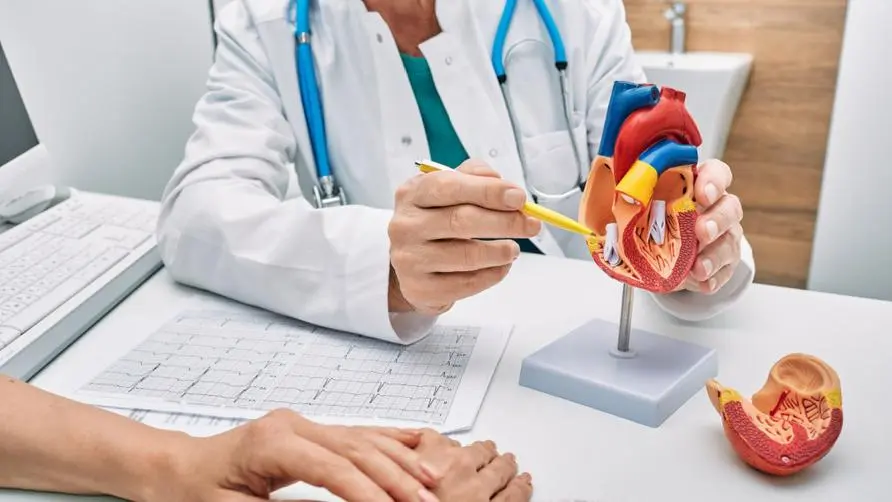
A cardiac ultrasound is a test that uses sound waves to produce real-time images of the heart. This test is mainly used to evaluate the function of the heart and valves. Cardiac ultrasound has a wide range of uses. In addition to being able to observe the patient’s heart structure, such as whether there are blood clots or thrombi in the heart chambers, the fluid accumulated around the heart, and the shape of the heart valves, cardiac ultrasound has some advantages that cannot be achieved by other imaging examinations. First of all, there is the radiation dose. There is no problem of radiation exposure in cardiac ultrasound, and there is no need to use a contrast agent. Even if a contrast agent is used, it is a contrast agent that does not damage liver and kidney function, and the risk and burden to the patient is very small.
The second is the dynamic assessment of cardiac function. General imaging examinations such as computed tomography and magnetic resonance imaging are relatively static examinations. It is difficult for the above examinations to see the dynamic changes of the heart in real time, such as the moment of valve opening and closing, heart contraction and Dynamic images of diastole, but these are the strengths of cardiac ultrasound.
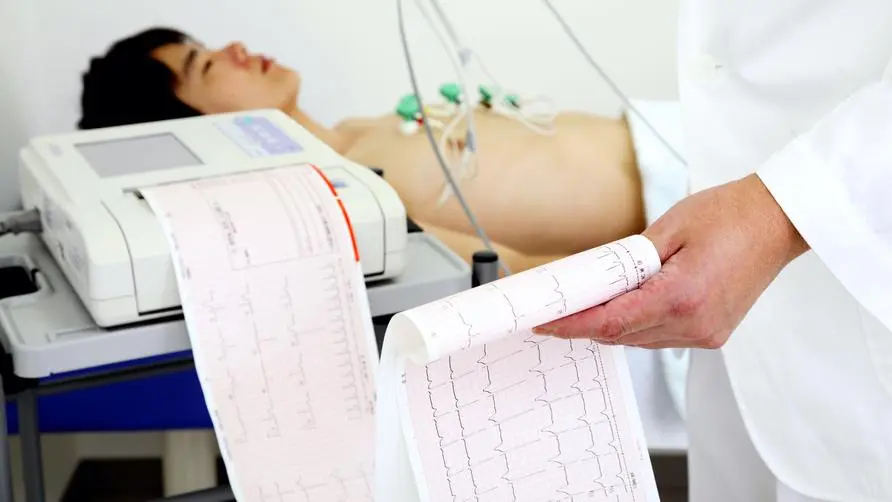
However, when testing for coronary artery disease, ultrasound is slightly less effective in assessing ischemia. Generally, there are two traditional non-invasive tests to evaluate myocardial ischemia, one is exercise electrocardiogram, and the other is nuclear medicine scan. The advantage of exercise electrocardiogram is that if the patient exercises, if there are any abnormalities on the electrocardiogram, it will show up.
Although exercise ECG is not 100% accurate, it is often used as an important evaluation before cardiac catheterization due to its convenience, no radiation, and no use of contrast agents. However, exercise electrocardiography can only tell doctors that there is ischemia in the myocardium, but it does not show which myocardial area is ischemic, and it is impossible to predict which blood vessels are narrowed. Cardiac nuclear medicine examination can make up for this shortcoming. In addition, drugs are used to simulate ischemia. Patients can know the extent of ischemia without running or cycling. For some groups with limited mobility, nuclear medicine examination is more suitable. However, this examination will use some drugs, and there are problems with nuclear radiation, which will have an impact on people around you, such as pregnant women.
Deformation analysis and electrocardiogram complement each other to make pre-cardiac surgery examinations more accurate.
Based on the above problems, without the use of drugs or radiation, the local and global contraction changes of the myocardium at rest and during exercise were compared using cardiac ultrasound. Although load-type ultrasound can show morphological abnormalities during movement, it is sometimes difficult to distinguish whether it is a real abnormality and the limits of the abnormality by naked eye observation. Therefore, cardiac ultrasound has also developed deformation analysis technology to visualize the location of myocardial ischemia.
The so-called deformation analysis technology uses software to analyze the position changes of the bright spots displayed on the myocardium during cardiac systole and diastole under ultrasound. The test is used to screen and track patients receiving cardiotoxic drugs during cancer treatment, to evaluate patients receiving chest radiation therapy, and to evaluate and monitor heart disease such as myocardial disease (e.g., hypertrophic, ischemic myocardial disease). .
Generally speaking, deformation analysis divides three sections of the left ventricle into 16 to 18 regions, and then reconstructs a circular chart of myocardial blood flow distribution, which is the so-called bull’s eye diagram. Based on the interpretation of the bull’s eye diagram, the extent of hypoxia in the patient’s heart muscle can be assessed and blocked blood vessels can be predicted. Studies have shown that stress-type ultrasound plus deformation analysis and nuclear medicine scans are equally accurate when compared with cardiac catheterization results.
In addition, when some patients have unexplained dyspnea, but various cardiac and respiratory system tests cannot find the cause, stress ultrasound may be used to detect other underlying diseases, such as diastole during exercise. Functional abnormalities or changes in mitral valve regurgitation, etc.
Stress-type ultrasound is developing rapidly and can complement other examinations to provide patients with more accurate examination quality before invasive surgery.


